Apr. 2, 2015

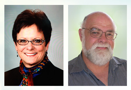
This year, ASRT members Gerald Conlogue, M.H.S., R.T.(R)(CT)(MR) and Carol Mount, B.S., R.T.(R)(M) received the International Speakers Exchange Award to present at the 2015 United Kingdom Radiological Congress and the 73rd CAMRT Annual General Conference, respectively.
In exchange, CAMRT is sponsoring Ashley A. Belbeck Ayume, B.Sc., MRT(T), to present at the upcoming ASRT Radiation Therapy Conference in September, and SOR is sponsoring James Stirling, M.Sc. in MRI, DCR, DMS, Radiographer Research Fellow, to present during the ASRT@RSNA sessions in December.
Learn more about the International Speakers Exchange Award.
About Carol Mount
Carol’s topic for the 2015 CAMRT Annual General Conference in Montreal, Canada, is "3-D Printing: The Next Technological Revolution in Radiology." Her presentation will provide a brief history and technical introduction to 3-D modeling, give an overview of Mayo Clinic’s 3-D modeling lab workflow, and depict 3-D models and the role they have played in surgical planning and other uses within the medical field.
Carol began her career at Mayo Clinic in 1971. Since that time she has held numerous positions ranging from staff technologist to supervisor of breast imaging and Intervention and, most recently, has acted as the supervisor of the Anatomic Modeling Unit and Coordinator of the Radiology Career Development Program. Over the span of her career her assignments have included educational, technical, quality and managerial duties.
Since 1992 she has published and presented numerous articles, lectures and posters covering topics ranging from mammography, image optimization and workflow optimization to ionizing radiation quality control. Her academic work has awarded her with the title of Assistant Professor of Radiology at Mayo Medical School. Her current focus has been on continuous improvement initiatives, and she recently achieved Gold Certification through Mayo Clinic’s Quality Academy.
Carol recently retired, but will remain active and involved in her radiology career. Carol believes everyone should take full advantage of every opportunity that is presented to them — most of the time, these turn out to be life-changing events!
About Gerald Conlogue
Gerald’s topic for the 2015 UKRC in Liverpool, England, is "Integrating Radiographers Onto the Forensic Team: A Decade at the Office of the Chief Medical Examiner for the State of Connecticut."
Gerald Conlogue is a professor of Diagnostic Imaging at Quinnipiac University. For over 40 years his research interests have focused on applying medical imaging techniques to nonmedical areas such as art conservation, wildlife management, archaeology, anthropology and forensics.
In 1999, Conlogue and Ron Beckett, emeritus professor of Health Sciences, founded the Bioanthropology Research Institute at Quinnipiac. Conlogue’s work with mummified remains led to appearances in several Discovery, History and Learning Channel productions in 2000. From 2001 to 2003 he and Beckett co-hosted "The Mummy Road Show" on the National Geographic Channel. During the series, they traveled to South America, Europe and Asia, employing imaging techniques to reveal clues to tell each mummy’s life story. In 2005, they published Mummy Dearest, a behind-the-scenes look and in-depth account of their experiences producing the series. Four years later, they published Paleoimaging: Field Applications for Cultural Remains and Artifacts, detailing the techniques and procedures they developed over the past 12 years.
For the past decade, Conlogue has taught an elective forensic imaging course for diagnostic imaging students and interested radiographers. Since 2002, he and his students have volunteered to radiograph cases at the Office of the Chief Medical Examiner in Farmington, Connecticut. In the 2010 publication Brogdon’s Forensic Radiography, he has a chapter that considers imaging mummified remains, and he was a contributing author on two chapters regarding imaging in the field and in the medical examiner’s facility.